Thayer County Health Services offers an extensive range of diagnostic imaging, using state-of-the-art imaging equipment. Over the years, the radiology department has grown steadily, committed to keeping pace with technological advances and our community’s needs.
THE RADIOLOGY DEPARTMENT PROVIDES:
Diagnostic X-ray examinations
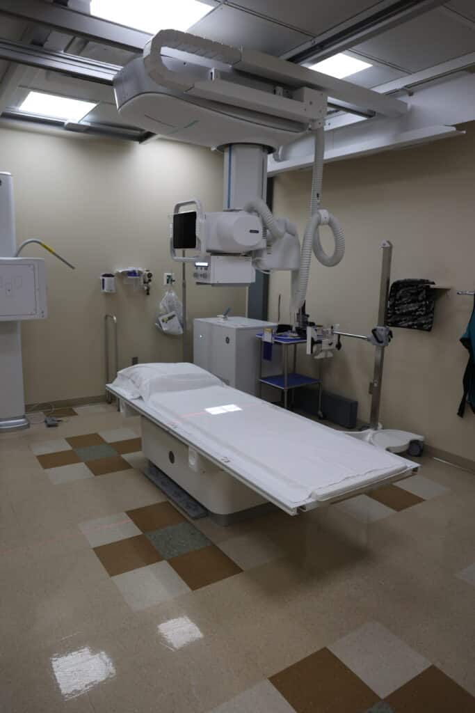
A diagnostic x-ray examination uses electromagnetic radiation to create images of the inside of your body to help doctors diagnose injuries or illnesses. It is a fast, non-invasive test used to detect conditions like broken bones, pneumonia, and tumors by showing different tissues and structures as varying shades of gray. Bones appear white because they block the radiation, while air in the lungs appears black.
Fluoroscopy and Barium studies
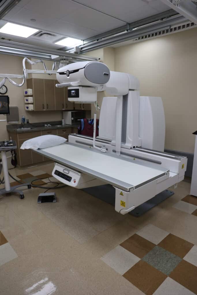
Fluoroscopy is an imaging technique that uses X-rays to create real-time, moving images of internal body structures, allowing doctors to see how organs and systems are functioning. A barium study is a type of fluoroscopy that uses a contrast agent like barium to make the gastrointestinal (GI) tract visible. Common barium studies include upper GI series and a barium swallow, which help diagnose issues like ulcers, strictures, or tumors in the esophagus, stomach, and small intestine.
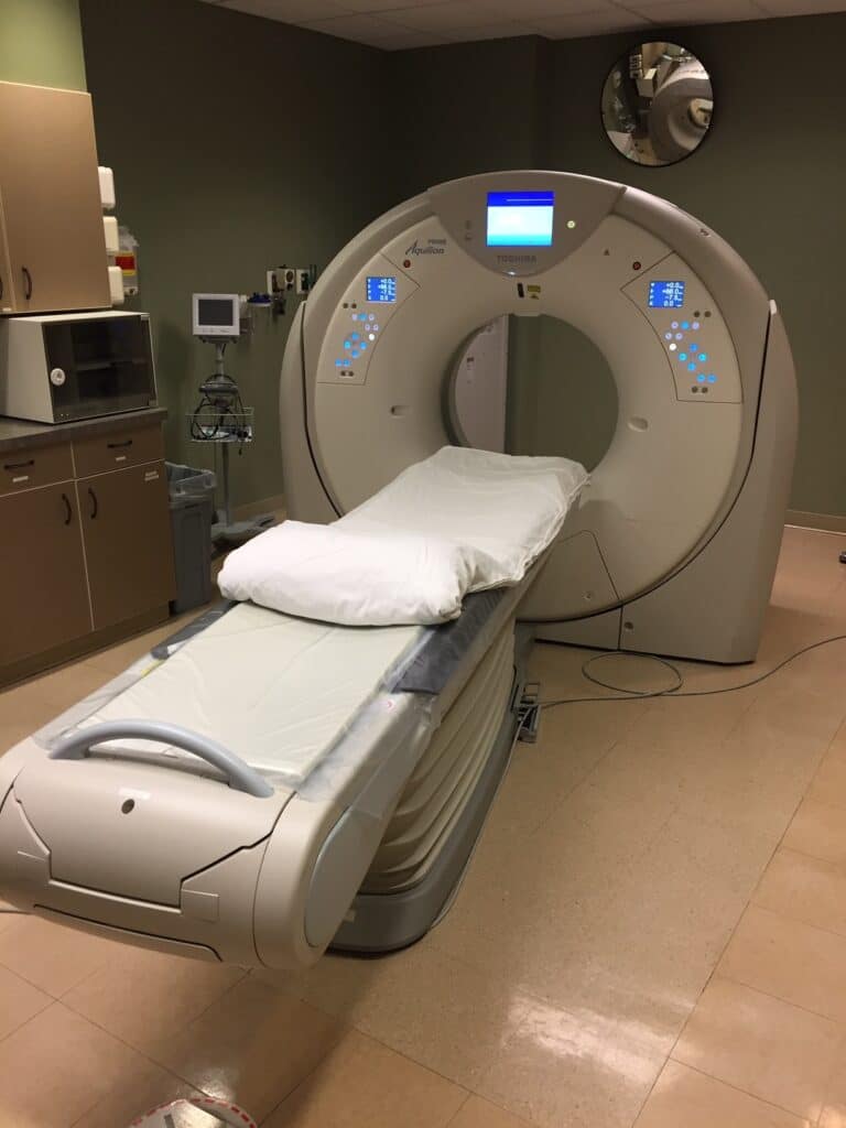
Computed Tomography (CT)
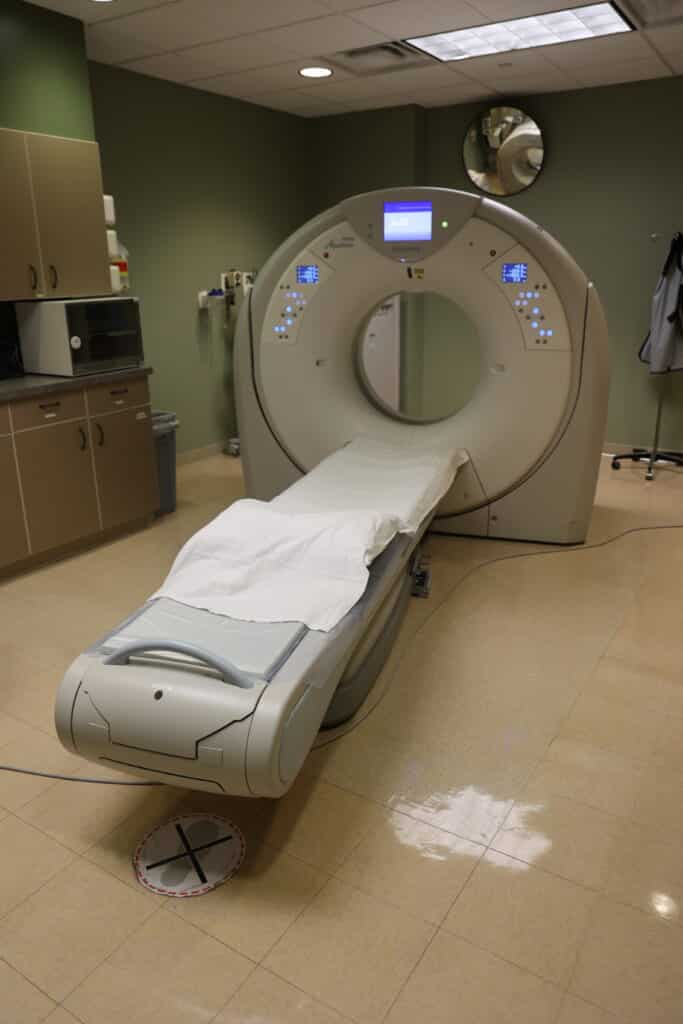
A CT (computed tomography) scan uses a series of X-rays and a computer to create detailed, cross-sectional images of the inside of the body, providing more clarity than a conventional X-ray. These images are often used to diagnose disease, plan treatment, and moniter how well a treatment is working by creating both 2D and 3D views of organs and tissues.
Magnetic Resonance Imaging (MRI)
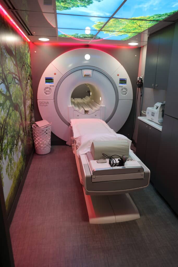
Magnetic Resonance Imaging (MRI) is a medical imaging technique that uses strong magnets and radio waves to create detailed, 3D images of organs, soft tissues, and bones. It does not use ionizing radiation, making it a safer option for frequent scans compared to X-rays or CT scans. MRI is used to diagnose and monitor conditions affecting the brain, spinal cord, nerves, lymph nodes, and other parts of the body, such as tumors, infections, and heart disease.
Bone Densitometry (DEXA)
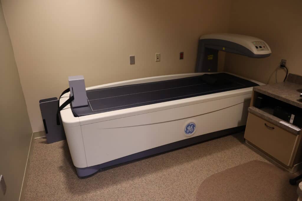
A DEXA scan is a quick, painless X-ray-based test that uses low-dose radiation to measure bone density and assess bone mineral content. It is the established standard for diagnosing osteoporosis and osteopenia (low bone mass) and for evaluating an individual’s risk of bone fractures. The test typically focuses on the spine and hips and is also used for body composition analysis, measuring fat, muscle, and bone mass.
Ultrasound
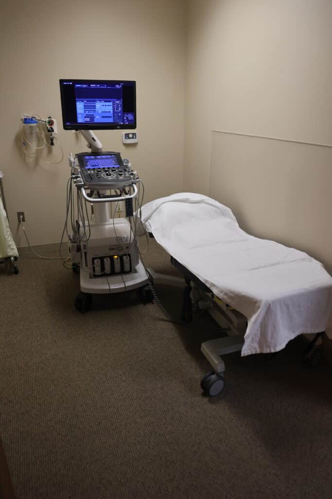
Ultrasound is a non-invasive imaging method that uses high-frequency sound waves to create real-time imaging of internal body structures. A transducer, or wand, is moved over the skin, sending sound waves into the body that bounce off tissues and return as echoes. A computer processes these echoes to produce an image, allowing medical professionals to examine organs, muscles, blood vessels, and more, and monitor fetal development.
3D Mammography
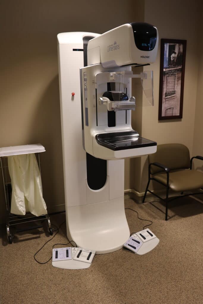
A 3D Mammogram, also known as digital breast tomosynthesis (DBT), is an advanced breast imaging technique that uses an X-ray machine to take multiple images of the breast from different angles. A computer then reconstructs these images into a 3D picture, allowing radiologists to examine the breast tissue into thin slices. This provides a more detailed view compared to a traditional 2D Mammogram, which can help improve the accuracy of detecting breast cancer, especially in women with dense breast tissue.
Nuclear Medicine
Nuclear Medicine is a medical specialty that uses small amounts of radioactive materials, called radiopharmaceuticals, to diagnose and treat diseases. It differs from other imaging techniques like X-rays or CT scans because it shows how organs and tissues are functioning, rather than just their structure. These radiopharmaceuticals are introduced into the body via injection, swallowed, or inhaled, and a special camera detects the radiation they emit to create images of the body’s internal processes.
Positron Emission Tomography (PET)
Positron Emission Tomography (PET) is a noninvasive imaging technique that uses a radioactive tracer to show how organs and tissues are working at a molecular level. A radioactive substance is injected into the bloodstream, and a doughnut-shaped scanner detects the positrons the tracer emits to create detailed 3D images of what’s happening inside the body. This allows doctors to assess blood flow, metabolic activity, and other biological processes in organ like the brain and heart, and to diagnose conditions such as cancer, Alzheimer’s, and heart disease.
Low-Dose CT Lung Screening
A Low-Dose CT (LDCT) Lung Screening is a quick, non-invasive imaging test that uses a small amount of radiation to create detailed, 3D images of the lungs. The goal is to detect lung cancer in its earliest stages, when it is most treatable and a cure is more likely.
For more information or to schedule an appointment, please call 402-768-4645.